
Radiological images are an essential part of the diagnosis process, and professionals need the best solutions for taking, storing, optimizing, and sharing those images across departments. That’s where PACS comes in – a system initially discussed in the 80s that has become an essential part of radiology departments worldwide for its many applications.
What does PACS stand for?
PACS stands for Picture Archiving and Communication Systems.
What is PACS, and how is it used in radiology?
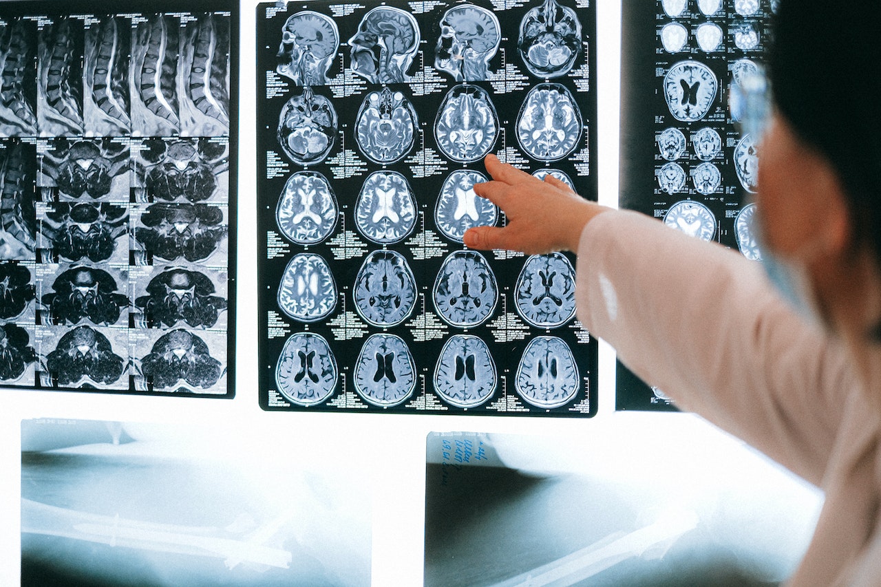
PACS are comprehensive computer systems that store and distribute 2D and 3D medical images for healthcare organizations. They provide an economical solution to the logistical realities of digitizing, printing, storing, retrieving, and sharing medical images across multiple devices, eliminating the need to manually handle X-ray films and medical reports.
It can handle images from various medical imaging instruments, including:
- Ultrasound
- Magnetic resonance
- Positron emission tomography
- Computed tomography scans
Radiology departments must store and sometimes share X-ray images and patient reports to maintain the department’s histories. PACS offers them centralized image storage, sharing, and appointment scheduling solution that significantly simplifies and organizes their data.
PACS can also be seen as a means for collaboration between multiple radiology departments.
If a patient undergoes several radiological investigations at separate locations, they may need to be accessed by multiple radiologists and physicians. PACS makes this collaboration possible by allowing them to transfer images while maintaining the original quality and staying HIPAA compliant.
What image types can PACS handle?
As mentioned before, PACS can handle multiple medical image types. Some of the most common supported image types include:
- Ultrasound (US)
- Magnetic resonance (MR)
- Nuclear Medicine imaging
- Positron emission tomography (PET)
- Computed tomography (CT)
- Endoscopy (ES)
- Mammograms (MG)
- Digital radiography (DR)
- Phosphor plate radiography
- Histopathology
- Ophthalmology
The image format shared by all PACS vendors is Digital Imaging and Communications in Medicine (DICOM), the standard for communicating and managing medical imaging information and related data. DICOM incorporates the standards for all these different image types and constantly includes new ones as they are developed.
PACS can also handle scanned documents and medical reports with PDF files.
What are the four components of PACS?
PACS consists of four major components.
1. Image acquisition devices such as computed tomography and magnetic resonance imaging
Imaging acquisition devices are composed of imaging modalities and acquisition gateway computers. These devices capture the image types PACS is designed to handle.
These devices are connected to the PACS server via acquisition gateway computers. Their main functions are acquiring images, converting them from their original formats to the PACS standard format (DICOM), and processing them for optimization.
2. A secured network for the transmission of patient information
A secure network is necessary to move medical data between PACS components, other systems, and remote locations. However, because PACS deal with medical information, they must provide a secured network to remain HIPAA compliant.
There are three main types of networks used to transfer radiological data:
- LAN networks are used within one medical department to link imaging devices, archive and data storage, and the display workstations
- WAN networks are used to connect different departments in a hospital and connect devices at large distances from each other
- Tele-radiology networks used to transfer medical data to remote hospitals in the region
3. Workstations (WSs) for interpreting and reviewing images
The display WSs are essentially personal computers used to analyze and review images. They are the hardware component that replaces the Alternator or the manual lightbox of the radiology system.
WSs are essential for making a primary diagnosis and are also named “diagnostic WSs.” They contain local storage databases, network connections for communications, resource management, displays, and processing software.
4. Archives for the storage and retrieval of images and reports
PACS servers archive all the patient’s data and imaging examinations, sent by the acquisition gateway computers and from HIS/RIS systems.
PACS servers have two main components: storage media (database) and archive systems. The archive systems operate on two levels: short and long-term archiving. Images from the short-term level take 2 seconds to retrieve, while images from the long-term level take 3 minutes.
What are the main uses and applications of PACS?
Radiology departments rely heavily on PACS for a myriad of uses and applications, including:
- Replace hard copies: PACS acts as a digital storage system that solves the logistical difficulties of storing physical copies, taking advantage of the decreasing cost of digital storage.
- Provide remote access: it allows practitioners to access accurate, optimized images across long distances for diagnosis and education purposes.
- Integrate electronic images: PACS integrates with other medical automation services to maximize effectiveness. These services commonly include Hospital Information systems (HIS), Electronic Medical Records (EMR), Practice Management Software, and Radiology Information Systems (RIS).
- Improve radiology workflow management: PACS is used mainly by radiology personnel to manage the workflow, but its applications are expanding to other fields.
What are the benefits and advantages of PACS?
PACS are extremely useful, and that’s why they’ve become an essential part of so many radiology departments around the world.
Benefits for healthcare organizations and radiology teams:
- Easily available images
- Better processing with multiple copies of the same image
- Image optimization
- Faster and better reporting
- Image sharing between various locations
- Digitization and archival of older films
- 2D and 3D image archival and processing
- Improved diagnosis due to higher-quality images
- Efficient data management
- Provides a chronological view of a patient’s radiology history
- Speeds up workflow
- Provides remote access
- Improved communication between doctors and hospital administration
- Potential applications in cardiology, oncology, pathology, dermatology, and nuclear medicine imaging
Benefits for patients:
- Reduced need to retake images and reexaminations leads to less radiation exposure.
- Faster, better diagnosis leads to better treatment
- More convenient. Patients don’t need to store images taken at different locations physically
What are the benefits of combining PACS and RIS?
PACS is designed to integrate with other automated technologies related to healthcare, such as Radiology Information System (RIS) and Hospital Information System (HIS). However, there are differences that are important to understand. The benefits of integrating PACS with RIS include:
- Richer patient information
- Increased consistency, accuracy, and availability of information
- The history and appointment information from RIS compliments PACS’s high-quality images and accessibility
- Less redundancy and human errors
- Faster access
- Enterprise-grade solutions can synchronize data and image management from different systems.
PACS are an integrated solution your radiology department needs
PACS is a highly robust data and image management system made with the requirements of radiology departments. Applied correctly, it can increase productivity and provide long-term image storage and retrieval solutions.
Radiology departments and other medical teams rely more on PACS to ensure they’re at the forefront of technology to maximize time and resources to provide a better experience for patients and their medical team.
Request a free PACS consultation from Nuvodia today! We’ll better understand your current healthcare imaging needs, review current technologies, and explain our current solution offerings to find the best-suited one for your organization.